ECG #3: Wraparound STEMI
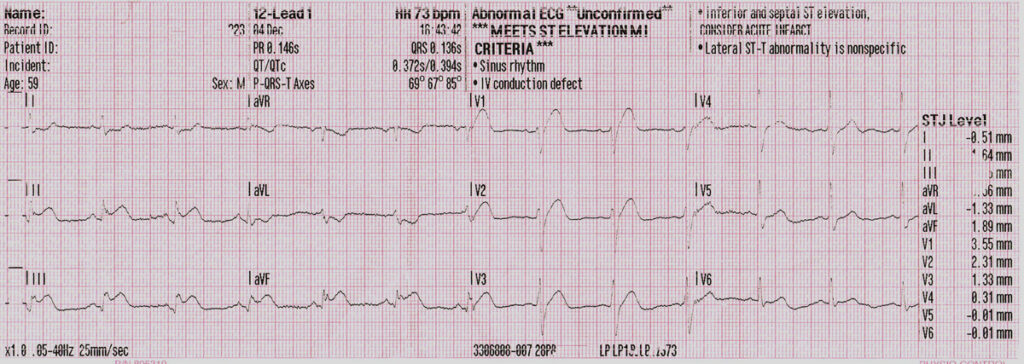
Here the patient was complaining of a sudden onset of central chest pain at rest, late in the afternoon on a Saturday. The leads went straight on and out came an obvious STEMI.
The ECG seems to show it’s both an inferior and anterior regions, or RCA and LCA vessels, that have been occluded at once! Maybe not so obvious in the anterior leads but the morphology of V2-V3 and the elevation in V1, it’s obvious that the septal region, as well as the inferiior region, is involved for sure.
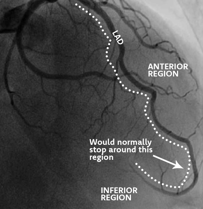
Uniquely, some patients can have a longer LAD vessel that travels down the anterior and wrapsaround the inferior of the heart finally finishing either on the bottom or posterior side of the heart. And thus inferior elevation is seen on the ECG.
According to both Kobayashi et al. (2015) and Tavakoli et al. (2021) patients with this type of STEMI typically have a worser outcome, logically as more regions of myocardial territory are insulted.
In this instance the patient remained stable with an LAD stent and a positive outcome at the cath lab.
References
Kobayashi, N. et al. (2015) “Usefulness of the left anterior descending artery wrapping around the left ventricular apex to predict adverse clinical outcomes in patients with anterior wall st-segment elevation myocardial infarction (an infuse-ami substudy),” The American Journal of Cardiology, 115(10), pp. 1389–1395. Available at: https://doi.org/10.1016/j.amjcard.2015.02.034.
Tavakoli, M. et al. (2021) “The comparing of short clinical cardiovascular outcomes with wraparound and nonwraparound left anterior descending artery in patients with anterior ST-segment elevation myocardial infarction,” Heart Views, 22(3), p. 184. Available at: https://doi.org/10.4103/heartviews.heartviews_216_20.