Tough Dysrhythmias (Amal Mattu, MD)

These are notes highlighting important points made whilst I was watching The Centre for Medicla. Education videos on Tough Dysrhythmias by Amal Mattu, MD, the legend.
Starting from sinus rhythm which is defined as:
- Upright P waves in I, II, III, aVF
- inverted in aVR.
V1 is the money lead in which to view the P waves as it sits directly over the R atrium.
Bradycardias
HR 40-60 = Junctional Bradycardia. Narrow. No p-waves as they are usually buried in the QRS.
HR 20-60 = Ventricular Escape Rhythm / Idioventricular Escape Rhythm. Wide.
AV Blocks
1st Degree
PR interval longer than 0.20. No treatment as it’s a normal part of aging. Can be seen in things like HyperK+, but you don’t treat the rhythm (1st degree) you treat the cause (tooooo much potassium).
2nd Degree Type I
aka Mobitz I aka Wenckebach. P’s on the run, Mobitz Type 1.
2nd Degree Type II
P’s stay true, Mobitz Type II. If you have 3p’s for every 2 QRS’s this is 3:2 bloc. If you have 2:1 you may not know if it’s even type 1 or 2 as there’s no 2nd pr for comparison with he first.
3rd Degree
Random PR interval. Lots of P’s.
Tachycardias
We need to know three things:
- Wide or Narrow?
- Regular or Irregular?
- Is there Atrial Activity (aka p or flutter waves?)
Narrow & Regular
Sinus Tachycardia: Most common. Never shock it. There’s a P for every QRS. The P is sometimes half-buried in the T wave appearing like a ‘camel-hump’.
Up to 220HR minus age. So a baby can be 2 and have a heart rate of up to 218 and still be called sinus tachycardia.
Supraventricular Tachycardia
Not ‘superventricular’. P’s are normally buried or follow the QRS aka a retrograde P wave, one that pops out after the QRS.
Atrial Flutter (2:1)
Normally 2 P waves for every QRS. Solid regular conduction.
Narrow & Irregular
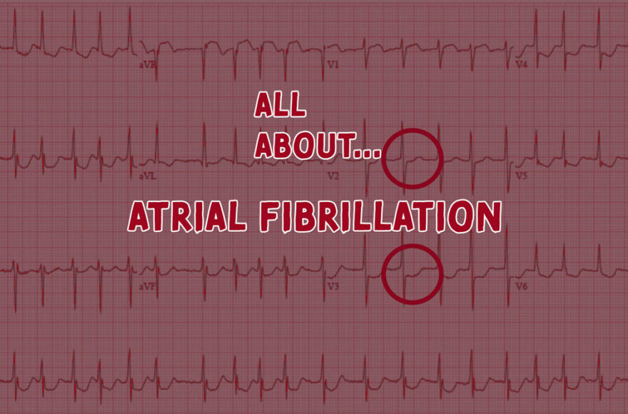
Atrial Fibrillation
The old classic. Differing R-wave heights. Super fast is offically called Atrial Fibrillation wih Rapid Ventricular Rate (AF+RVR)
Atrial Flutter with Variable Conduction
Instead of regular like above, it conducts in various ratios: 2:1, 3:1, 2:1, 5:1, 4:1, 5:1, 2:1….
Multifocal Atrial Tachycardia (MAT)
These have more than three morphologies of P wave (inverted, biphasic, flat, tall). Never shock this badboy. Normally associated with pulmonary diseases like COPD, asthma, peumonia).
Wide and Irregular
Sinus Tachycardia with Abberent Conduction
This is tachycardia with a bundle branch block (BBB), normally P-wave present and wide QRS.
Ventricular Fibrillation
- P-waves hidden / occasionally seen
- AA/V disassociation
- Must be >120/130. If not, maybe a mimic:
- HyperK+
- Tricyclic Acid OD
- Accerlated Idioventricular Rhythm
Supraventricular Tachycardia with Bundle Branch Block
P waves are often hidden
Atrial Fibrillation with Bundle Branch Block
Ths is rare to see >200HR. Also have identical QRS.
Atrial Fibrillation and Wolff-Parkinson White
Rate is normally around 300HR. QRS would vary morphology. Appears regular at very rapid rates.
Other Tachycardias
Polymorphic VT
- Regular
- Rapid
- Distinction between Poly VT and TdeP n ot made by looking at QRS complexes during arrhythmia.
- Shock it!
- Polymorphic VT
- No prolonged QT prior
- Caused by Acute Cardiac Ischaemia
- Treated with Amioderone / Procainamide
- Torsades de Pointe
- Prolonged QT prior
- Caused by Drugs or Hypoelectrolytes
- Treated With Magnesium, Amioderone, Procainamide
Accelerated Idioventricular Escape Rhythm (AIVR)
HR 40-120, normally a reperfusion arrhythmia that looks like VT and lasts 5-10 seconds. Seen lots in cath labs when restoring blood flow to vessels.
4 Cardinal Sins of Instability
Hypotension
Confusion
Ischaemic Chest Pain
Acute Heart Failure


